
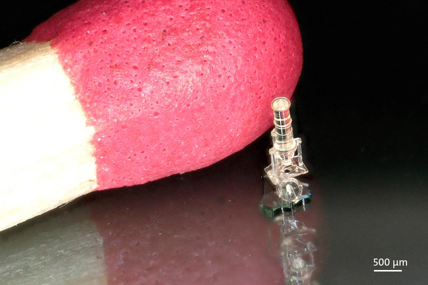
A research group at TU Wien has used ultra-precise 3D printing to grow replacement cartilage that, in a rabbit knee model, closed defects to roughly 91% of natural tissue thickness within 12 weeks — a result reported by VoxelMatters as part of a collaboration between TU Wien, the Ludwig Boltzmann Institute for Traumatology (AUVA), and the Medical University of Vienna. The team, led by Prof. Aleksandra Ovsianikov with PhD student Oliver Kopinski-Grünwald among those working on the project, printed the scaffolds using two-photon polymerization and a biodegradable resin from Belgian materials company BIO INX.
Cartilage is a notoriously bad self-healer. It has no blood supply, so once it's damaged — by injury, wear, or osteoarthritis — the body has almost no mechanism to rebuild it. That's the gap this project is aimed at: not a bigger, easier print, but a smaller and more precise one, engineered to give cells a scaffold that behaves enough like real cartilage architecture that the body accepts it as a starting point for regrowth.
The Print: Small, Precise, and Loaded With Living Cells
The core fabrication method here isn't FDM or even standard resin DLP/SLA — it's two-photon polymerization (2PP), also called multiphoton lithography. Instead of curing an entire resin layer at once with a UV lamp or projector, 2PP uses a tightly focused femtosecond laser that only solidifies resin at the exact focal point, where two photons arrive simultaneously with enough combined energy to trigger polymerization. That lets the printer build features far below what a single-photon process can resolve, tracing out fine three-dimensional lattice geometries voxel by voxel.
The TU Wien group used this precision to print microscaffolds just 0.3mm in diameter — structures sized to match the scale of a single defect site rather than a bulk implant. The resin itself, BIO INX's DEGRAD INX, is a biodegradable material formulated specifically to break down in the body over time, ideally on a timeline that hands off structural support to newly formed tissue as the scaffold dissolves.
Rather than printing an empty lattice and hoping cells migrate in on their own, the researchers loaded each scaffold with spheres of cartilage cells derived from stem cells — pre-formed clusters designed to behave like functional cartilage tissue from the start. The scaffold's job is largely mechanical and architectural: hold those cell spheres in the right three-dimensional arrangement, at the defect site, long enough for them to integrate and mature.
What Happened in the Rabbit Model
The constructs were implanted into an osteochondral-defect model in rabbit knees — a standard preclinical setup for testing cartilage repair, where a controlled defect is created down through the cartilage layer and into underlying bone. After 12 weeks, the regenerated tissue reached approximately 91% of natural cartilage thickness, with VoxelMatters' report describing the new tissue as integrating well with the surrounding native tissue. A separate item in 3DPrint.com's September 5 news briefs confirms the same collaborators, resin, scaffold diameter, and 12-week timeframe, and notes the implanted rabbits showed majorly improved cartilage regeneration compared to untreated control animals. Both reports describe this as a preclinical animal-model result, not a human trial — 91% thickness recovery and good tissue integration in rabbits is a meaningful data point on the way toward eventual clinical application, not a finished therapy. Cartilage regeneration research routinely runs through small-animal models like this before any larger-animal or human work is even attempted, and the distance between "worked in a rabbit knee" and "approved treatment" is typically measured in years.
Why Two-Photon Printing, Specifically
It's worth dwelling on why the team reached for one of the slowest, most expensive additive processes available rather than a conventional bioprinting extrusion setup. Cartilage's mechanical function comes largely from its microstructure — the fine-scale arrangement of its extracellular matrix — not just its bulk shape. A scaffold that's the right overall size but the wrong internal geometry at the tens-of-micron scale won't guide cell behavior correctly. Two-photon polymerization is one of the few fabrication methods that can hit that resolution in three dimensions while still working with a resin chemistry, like BIO INX's DEGRAD INX line, engineered for biocompatibility and controlled degradation. The tradeoff is throughput: 2PP builds voxel by voxel with a scanning laser, so it is inherently a small-part, high-value process, not something suited to printing sheets of scaffolds at volume. That tradeoff is presumably fine here — a 0.3mm implant destined for a single defect site doesn't need to be mass-manufactured the way a consumer part does; it needs to be right.
This is also why a multi-institution team was needed rather than a single lab working alone. TU Wien's role is the printing side — the laser hardware, the scanning strategy, and the process knowledge needed to turn a resin into a reliable microscaffold. The Ludwig Boltzmann Institute for Traumatology and the Medical University of Vienna bring the clinical and surgical expertise required to design a realistic defect model, implant the constructs, and evaluate the resulting tissue. BIO INX supplies the resin chemistry itself. None of those pieces alone gets a scaffold from a CAD file into a living rabbit knee and back out again as evaluable tissue; the collaboration structure reflects how specialized each stage of this kind of biofabrication work has become.
What It Means for Makers
This isn't a project anyone will replicate on a desktop resin printer — 2PP hardware occupies an entirely different tier from the Formlabs or Elegoo units in most maker workshops, and DEGRAD INX is a specialty biomedical resin, not a shelf item. But it's a useful data point for anyone tracking where photopolymer resin chemistry is headed. BIO INX has been positioning its DEGRAD INX line as an off-the-shelf biodegradable option for research groups building bioprinting and tissue-engineering workflows, rather than requiring every lab to formulate its own resin from scratch — the same commoditization pattern that's played out with engineering-grade FDM filaments and standard SLA resins over the past decade. As more validated, application-specific resins become available as ready-made products, the barrier to running serious biomedical printing research drops, even if the printers themselves stay in specialized labs. For makers with an interest in materials science, DEGRAD INX and results like this one are a signal of how far resin engineering has moved beyond "prints a rigid part" toward "prints a part that a body can safely absorb."
