
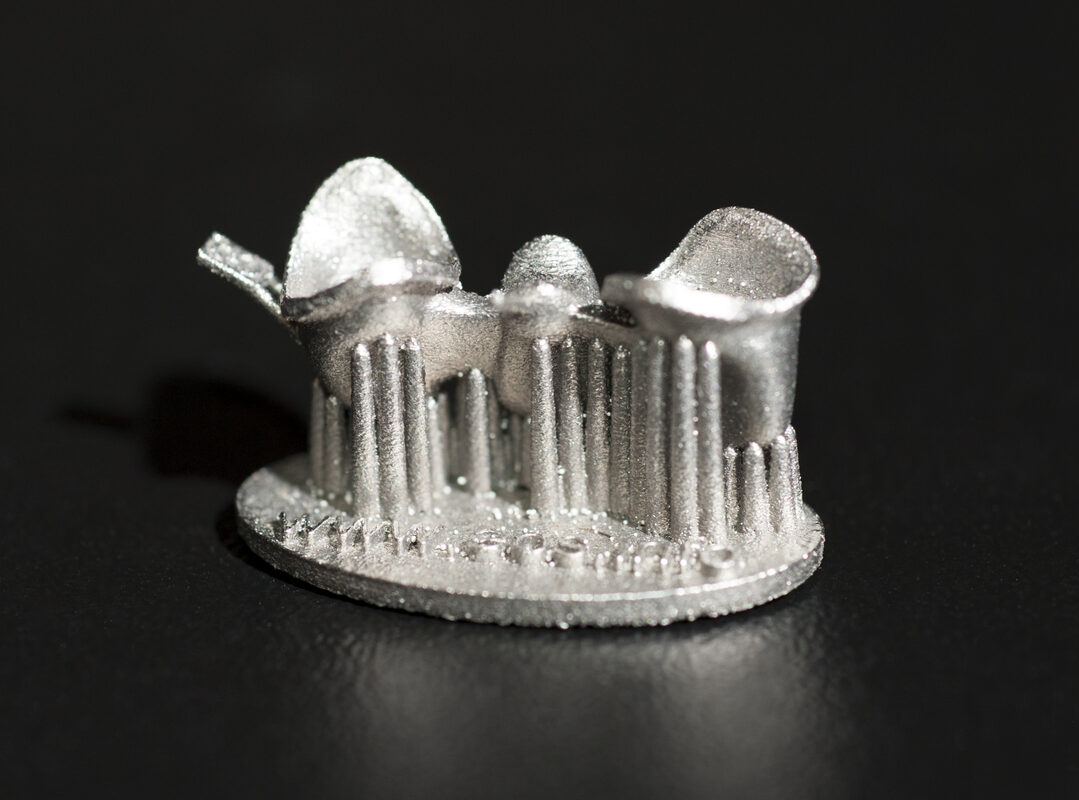
Dental students have long trained on models that look like teeth but don't behave like them — plastic or resin substitutes that hold their shape under a drill or elevator instead of chipping, cracking, and giving way the way real enamel and dentin do. A new Adelaide University-led project aims to close that gap by 3D printing dental replicas engineered to fracture like the genuine article. Announced August 18, 2026, the 18-month, AU$800,000 collaboration pairs Adelaide University with medical training device company Fusetec and the Additive Manufacturing Cooperative Research Centre (AMCRC), combining clinical imaging, digital modeling, and multi-material additive manufacturing to build patient-specific teeth, jawbone, and soft-tissue replicas.
The project is led by Associate Professor Ling Yin of Adelaide University's School of Electrical and Mechanical Engineering, working alongside the School of Dentistry and the Future Industries Institute. "Every patient presents differently, yet existing training models cannot accurately replicate the feel and behaviour of human dental tissue," Yin said. "By combining advanced clinical imaging with additive manufacturing, we can create dental models that better mimic real anatomy and help clinicians understand the forces involved in complex tooth removal procedures." The stated goal isn't just anatomical accuracy — training mannequins have offered that for decades — but replicating surgical response: how tissue actually gives way when a clinician applies force during procedures like wisdom tooth extraction.
Why Fracture Behavior Is the Hard Part
Anyone who has printed a mechanical part knows that geometry and material are two separate problems. A model can be dimensionally perfect and still behave nothing like the object it represents once load is applied — wrong stiffness, wrong failure mode, wrong feel under the tool. That's the specific problem this project targets. According to Manufacturers' Monthly's coverage, researchers will develop simulation tools to better understand fracture-force thresholds during surgery — essentially building a data-driven model of exactly how much force it takes to crack a tooth or section of jawbone, and in what pattern, before translating that into printable, multi-material geometry, in support of what both source articles describe as "safer, more predictable and less invasive clinical practice." That's a substantially harder target than the surface fidelity most dental training aids chase. A tooth isn't a single material — enamel, dentin, pulp, periodontal ligament, and bone all have different stiffness and fracture characteristics, and they're bonded together in a specific structure that determines how a crack propagates when an elevator or forceps applies torque during extraction. Reproducing that with a single-material print is essentially impossible; it requires multi-material additive manufacturing capable of varying mechanical properties within a single build, print zone by print zone, to approximate the layered structure of real oral anatomy.
The pipeline described combines clinical imaging — presumably CT or CBCT scans of real patient anatomy — with digital modeling to generate patient-specific geometry, then multi-material printing to realize it physically. That's consistent with how the broader dental and surgical training industry has been moving: away from generic, one-size-fits-all mannequins and toward models built from real patient data, which lets trainees rehearse on anatomy that resembles what they'll actually encounter in a specific procedure, including unusual root shapes or bone density.
The Backing: Fusetec and AMCRC
The project runs under the Additive Manufacturing Cooperative Research Centre, an industry-linked research vehicle that pairs university expertise with a commercial partner positioned to productize the output — here, Adelaide's mechanical engineering and dentistry schools working with Fusetec. AMCRC managing director Simon Marriott framed the project as a template for how the centre operates: "The opportunity starts with industry. Fusetec identified a clear clinical challenge and partnered with Adelaide University to develop an advanced manufacturing solution with strong commercial potential," he said. Neither source discloses AMCRC's total funding pool or partner count, so those figures are omitted here rather than estimated. Fusetec CEO Mark Roe framed the work as moving existing research toward a commercial product: "This collaboration allows us to turn years of research into a commercially viable product with real clinical value," Roe said. "We're developing dental models that don't just look like anatomically correct — they respond like real tissue during surgery." That framing points toward the eventual path for the work: a product line for dental schools and training programs, not just a research exercise.
Neither source publishes a technical specification sheet — no named printer, no disclosed material chemistry, no resolution figures — which is typical for an announcement at project kickoff rather than at results. What's confirmed is scope and structure: an 18-month timeline, an AU$800,000 budget, and a three-way collaboration between a university engineering/dentistry team, a training-device manufacturer, and a CRC funding and industry-network vehicle.
What It Means for Makers
This isn't a project makers will replicate on a desktop FDM printer, but it's a useful data point for anyone tracking where multi-material additive manufacturing is heading in medical and training applications. A few things worth noting:
Multi-material is the differentiator, not resolution. The interesting engineering problem here isn't printing a tooth-shaped object — plenty of consumer-grade resin printers can already do that with visually convincing results. It's printing an object where different regions have deliberately different mechanical properties that interact correctly under load. That's the frontier multi-material platforms (think Stratasys PolyJet-class machines or similar) are being pushed toward across medical, aerospace, and now training-device markets, and it's a capability gap that separates hobbyist and prosumer printing from the high-end multi-material systems this kind of project will likely require.
Patient-specific pipelines are becoming standard practice. The clinical-imaging-to-print workflow described here mirrors what's already common in patient-specific surgical guides, anatomical models for pre-op planning, and prosthetics — CT/CBCT scan in, segmented digital model, multi-material or single-material print out. Dental training replicas following the same pattern suggests this workflow is maturing from novelty to standard tooling across medical additive manufacturing.
Simulation-to-print feedback loops matter. Building fracture-force simulation tools alongside the physical models — rather than just eyeballing whether a printed tooth looks right — reflects a broader trend of pairing FEA-style mechanical simulation with additive manufacturing workflows to validate that a print will actually perform the way its geometry suggests, before it's produced.
For now, this is a funded 18-month research effort with a defined budget and named leads, not a shipping product. The value for the wider AM community is mostly directional: another concrete example of multi-material 3D printing being tasked with reproducing not just the shape of biological tissue, but its behavior under mechanical stress — a bar that's considerably higher than most training-model manufacturing has historically aimed for.
